山梨肺癌研究会会誌 第11巻1号 022-030(1998)
巨大肺嚢胞症術後に発生した肺癌の1例
古屋一茂、霜多 広、窪田健司、石原重樹、堤 正夫、大和庸次、椿原基史
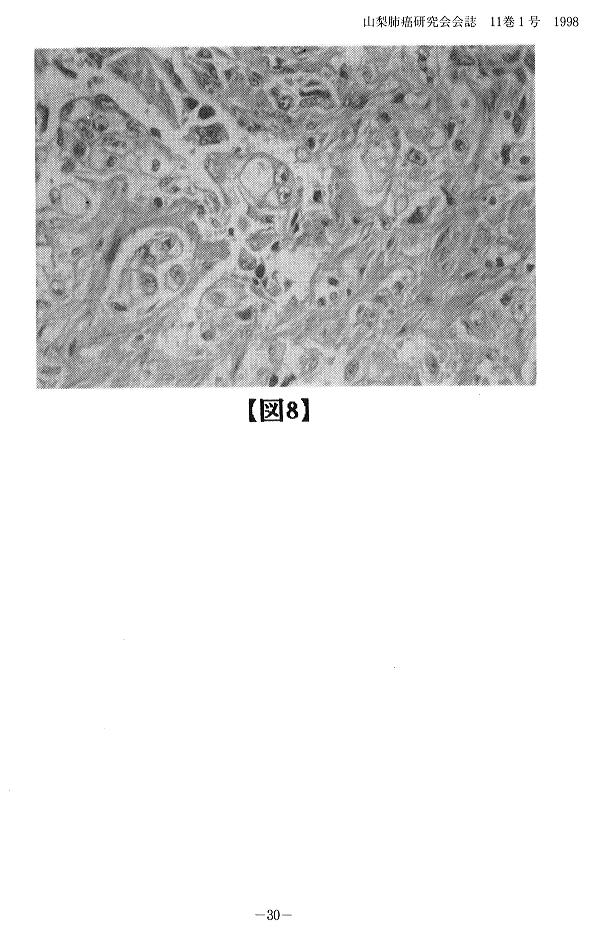
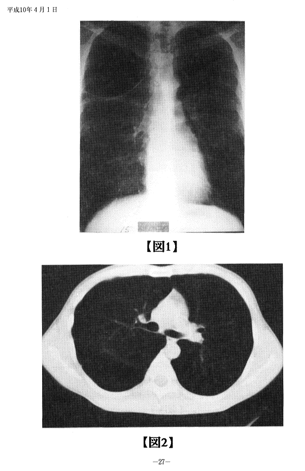
要旨:症例は41歳男性、巨大肺嚢胞症の診断平成7年8月22日右肺ブラ切除術を行った。切除ブラ組織に悪性病変部は存在しなかった。1年6カ月後に胸痛の訴えあり、平成9年2月の胸部X線写真上右上肺野に腫瘤陰影を認めた。肺癌の診断で同年4月8日右上葉切除術を施行した。切除標本で、腫瘍の占拠部位は、S1、S2で大きさは5.0X1.5X6.0cm。壁側胸膜への直接浸潤を認め、胸膜の断端は陽性で組織学的には大細胞癌であった。術後放射線治療を行ったが、術後6カ月後に死亡した。気腫性肺嚢胞症に肺癌が合併し易い事は以前から指摘されているが、術後経過観察中、残存した気腫性肺組織から発生した症例と考え報告した。
Key words:Giant emphysematous bullae、Lung cancer
A case of lung cancer originating at the site of previous bullectomy
Kazushige Furuya, Hiroshi Shimada, Kenji Kubota, Shigeki Ishihara, Masao Tutumi, Youji Yamato, Motofumi Tubakihara
A 41 year-old mas was admitted to our hospital with a complaint of chest pain, on the right. He had a past history of a giant bullae on the upper lobe of the right lung and had undergone bullectomy two years earlier. the histology of the specimen had proven to be negative for malignancy. But 18 months from then, he was found to have an abnormal shadow on the right upper lobe in chest X Ray. Subsequent investigations revealed that to be a mass, and he underwent a right upper lobectomy. Examination of the specimen confirmed the mass to be a tumor of 5.0X1.5X6.0 cms dimension, originating from the right upper lobe, with invasion into the parietal pleura, as the resected pleural margins were positive for malignancy. Histologically the tumor was a large cell carcinoma with mucin. In spite of postoperative chemotehrapy and radiation therapy, the patient died 6 months after the surgery.
many cases of lung cancer associated with giant emphysematous bullae have been reported in the japanese literature. Those cases including ours, insist the importance of through pre, and postoperative check-up of patients presenting with emphysematous bullae, having in mind, the possibility of a carcinoma behind them.
本文ページです。見たいページをクリックして下さい。(解像度が低いため、鮮明な画像が必要な場合は原本をご参照下さい)
p. 22 p. 23
p. 23 p. 24
p. 24 p. 25
p. 25 p. 26
p. 26
p. 27 p. 28
p. 28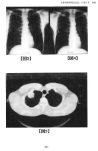 p. 29
p. 29 p. 30
p. 30
目次・Contentsに戻る
 p. 23
p. 23 p. 24
p. 24 p. 25
p. 25 p. 26
p. 26
 p. 28
p. 28 p. 29
p. 29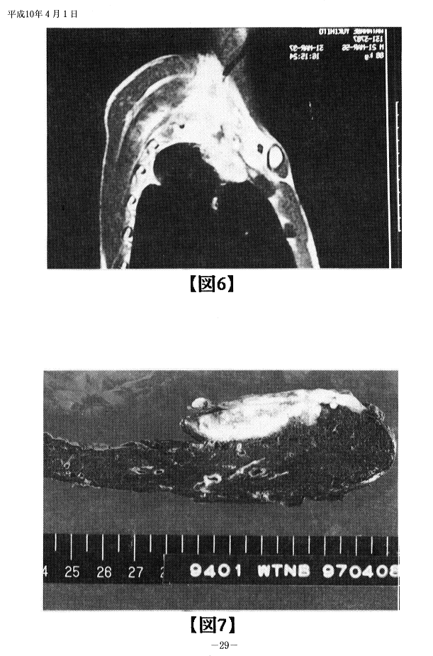 p. 30
p. 30