山梨医科大学紀要 第10巻,049-056(1993)
臓器移植と放射線医学
―米国 Pittsburgh 大学での経験―
大友 邦
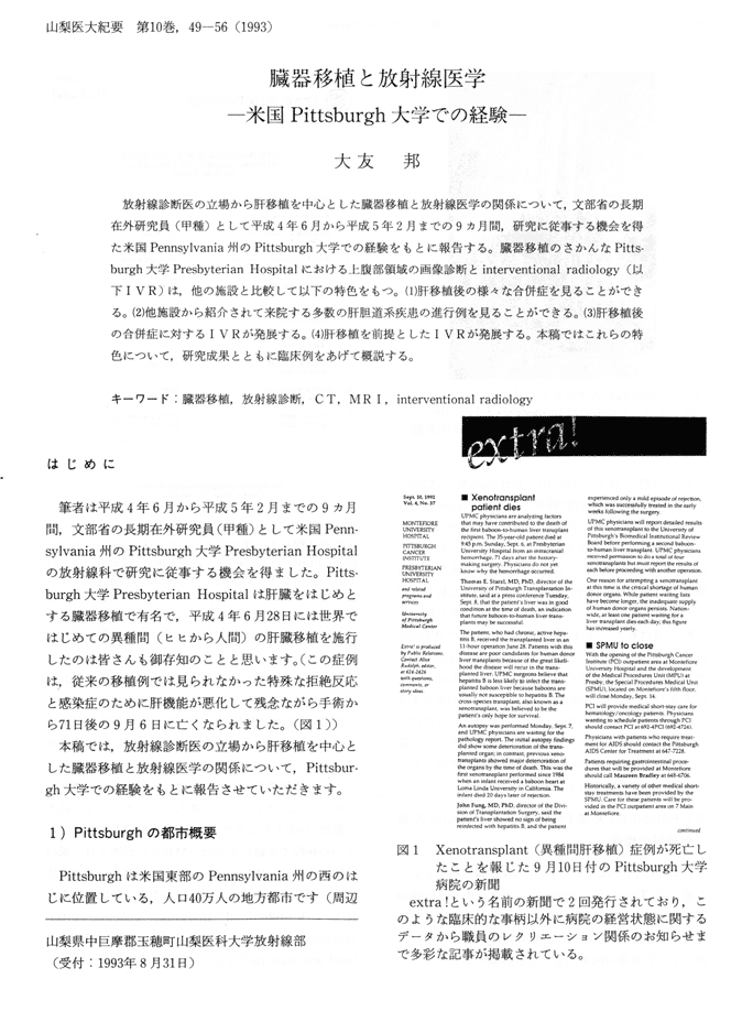
放射線診断医の立場から肝移植を中心とした臓器移植と放射線医学の関係について、文部省の長期在外研究員(甲種)として平成4年6月から平成5年2月までの9カ月間、研究に従事する機会を得た米国Pennsylvania州のPittsburgh大学での経験をもとに報告する。臓器移植のさかんなPittsburgh大学Presbyterian Hospitalにおける上腹部領域の画像診断とinterventional radiology(以下IVR)は、他の施設と比較して以下の特色をもつ。(1)肝移植後の様々な合併症を見ることができる。(2)他施設から紹介されて来院する多数の肝胆道系疾患の進行例を見ることができる。(3)肝移植後の合併症に対するIVRが発展する。(4)肝移植を前提としたIVRが発展する。本稿ではこれらの特色について、研究成果とともに臨床例をあげて概説する。
キーワード:臓器移植、放射線診断、CT、MRI、interventional radiology
CT and MR Imaging of Confluent Hepatic Fibrosis in Advanced Cirrhosis
Kuni OHTOMO
1) CT
To determine CT characteristics of confluent fibrosis complicating liver cirrhosis, CT scans from 420 cirrhotic patients without hepatic malignancy who underwent hepatic transplantation were correlated with specimens of freshly resected whole livers. In 59 of the 420 patients, 70 focal abnormalities corresponding to confluent fibrosis were revealed on CT. The lesions were characterized by shape: 49 lesions appeared wedge shape and radiated from the porta hepatis (involving the medial segment of the left lobe and/or the anterior segment of the right lobe in 44) ; eight lesions were peripheral and band-like ; 13 lesions were seen as total lobar or segmental fibrosis (right lobe-4, lateral segment of the left lobe-9). Associated volume loss is the affected regions were seen in 62 of the 70 lesions and was seen as retraction of the overlying hepatic capsule or total shrinkage of the segmental/lobar involvement. All 70 lesions were seen on CT as areas of lower attenuation than adjacent liver on non contrast CT and 51 of 64 lesions became areas of iso density or minimally lower attenuation on post contract CT. Greater enhancement than adjacent liver was demonstrated in eight and areas remained significantly lower in attenuation than liver in the other five. Our study shows that confluent fibrosis produces a characteristic appearance on CT scans and the recognition of its characteristics may help radiologists to differentiate it from hepatic neplasms in cirrhotic patients.
2) MRI
The value of MR imaging in the diagnosis of confluent fibrosis in advanced cirrhosis was assessed by pathologic features of l1 cirrhotic patients with confluent fibrosis. On T1-weighted spin echo (SE) images, 10 lesions were hypointense and one was isointense. All lesions were hyperintense on T2-weighted SE images (l1/1l) and on short T1 inversion recovery images (6/6). Greater enhancement than adjacent liver was not revealed in five lesions studied with dynamic MR imaging. Microscopic evaluation revealed fibrosis with prominent edema, which might explain the signal intensity of these lesions on MR images. MR imaging provided vseful morphologic information about confluent fibrosis, however confluent fibrosis could not be differentiated from hepatic neoplasms with signal intensities of MR images alone.
本文ページです。見たいページをクリックして下さい。(解像度が低いため、鮮明な画像が必要な場合は原本をご参照下さい)
p. 49 p. 50
p. 50 p. 51
p. 51 p. 52
p. 52 p. 53
p. 53
p. 54 p. 55
p. 55 p. 56
p. 56
目次・Contentsに戻る
 p. 50
p. 50 p. 51
p. 51 p. 52
p. 52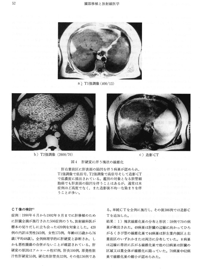 p. 53
p. 53
 p. 55
p. 55 p. 56
p. 56