山梨医科大学紀要 第2巻,001-007(1985)
腸管神経叢の骨格
モルモット小腸のS-100蛋白免疫陽性細胞に
関する研究から
小林 繁、鈴木道子、遠藤登代志
"腸管神経叢の骨格"を提唱し、その解剖について解説した。
モルモット小腸壁の各層の剥離標本を抗S-100蛋白血清をもちいたペルオキシダーゼ・抗ペルオキシダーゼ法で免疫染色し光学顕微鏡で観察した。免疫陽性染色は3次元的な広がりをもつ一群の細胞(S-100細胞)に限って特異的に認められた。この細胞系の立体構造のあらましは、ヘチマの果実から作られる束子をモデルにして記述できる。私たちはS-100細胞が腸管神経叢の骨格の主要部分を形作るとの観察に基づき、漿膜下層、外縦筋層、Auerbach神経叢、内輪筋層浅部、内輪筋層深部、粘膜下層特に Meissner神経叢、粘膜筋板および粘膜についてそれぞれに特有な骨組みのパターンを図説した。腸管神経叢の神経細胞の突起は、ぶどう蔓をからめるぶどう棚のように、Sー100細胞製の骨格がこれを支える。
キーワード:腸管神経叢、腸管神経系、Sー100蛋白、免疫組織化学、Auerbach神経叢、Meissner神経叢
On the Cellular Skeleton of the Enteric Nervous System
- An Immunocytochemical Study in the Guinea-Pig Small Intestine
Using an Anti-S-100 Protein Serum -
Shigeru KOBAYASHI, Michiko SUZUKI and Toyoshi ENDO
This is an essay on the three-dimensional structure of the enteric nervous system which is likened to that of a loofah. We investigated in light microscopy the whole-mount preparations of the layers of the guinea-pig small intestine by the peroxidase-antiperoxidase method using an antiserum to S-100 protein. There was a delicate network of the S-1OO protein immunopositive cel1s (S-1OO cells) extending in the subserous plexus, longitudinal muscle layer, Auerbach's plexus, circular muscle layer including the deep muscular plexus, submucous layer including the Meissner's plexus, lamina muscularis mucosae and lamina propria mucosae. We propose that the cellular network composed of the S-1OO cells forms the skeleton of the enteric nervous system. This network, as the trellis holding the grape-vines, provides a framework for supporting the processes of the enteric neurons.
本文ページです。見たいページをクリックして下さい。(解像度が低いため、鮮明な画像が必要な場合は原本をご参照下さい)
p. 1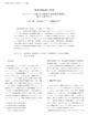 p. 2
p. 2 p. 3
p. 3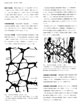 p. 4
p. 4 p. 5
p. 5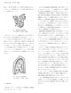
p. 6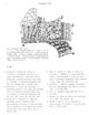 p. 7
p. 7
目次・Contentsに戻る
 p. 2
p. 2 p. 3
p. 3 p. 4
p. 4 p. 5
p. 5
 p. 7
p. 7