山梨医科大学紀要 第8巻,029-034(1991)
肝画像診断の最近の進歩
大友 邦、内山 暁
画像診断の一般の進歩を促す要因は、診断機器及び診断手技の改良と臨床医字の変遷進歩に分けることができる。診断機器の進歩として、超音波におけるドップラー法の導入、CTでは超高速CTの臨床応用とX線検出器の改良、MRIでは MR angiographyと MR spectroscopyの実用化と高速撮像法の進歩があげられる。また診断手技の工夫のうち肝臓を対象としたものでは、超音波と血管造影を組み合わせたCO2動注下の超音波検査、CTと血管造影を組み合わせた門脈造影下CT、ガイドワイヤーなどの改良により容易となった超選択的肝動脈造影がある。臨床医学の変遷は新たな疾患概念の導入と新たな治療法の導入に分けられ、肝臓については前者では肝細胞癌の前駆病変としての腺腫様過形成や早期肝癌、後者では肝移植がある。本稿ではこれらの様々の要因による肝疾患の画像診断の進歩について概説した。
キーワード:肝臓、画像診断、超音波検査、X線CT、MRI
Recent Advances of Diagnostic Imagings
of the Liver
Kuni OHTOMO, Gyo UCHIYAMA
Factors which stimulate advances of diagnostic imagings are divided into two groups. The first ones are improvements of the hardware and the software of the imaging modalities. They include Duplex and 2D Doppler Ultrasonography, ultrafast CT, MR angiography, MR spectroscopy and various fast scan techniques of MRI. CO2 ultrasonography and CT during arterial portography are also inventions of the software. Superselective hepatic arteriography is now easily and safely performed with torque-controllable guidewires. The second groups are newly introduced concepts and treatment of hepatic diseases. For example, the former includes adenomatous hyperplasia which is one of the precursors of hepatocellular carcinoma and the latter includes liver transplantation. In this article, we briefly review the recent advances of diagnostic imaging of the liver based upon the classification of the causative factors mentioned above.
本文ページです。見たいページをクリックして下さい。(解像度が低いため、鮮明な画像が必要な場合は原本をご参照下さい)
p. 29 p. 30
p. 30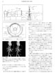 p. 31
p. 31 p. 32
p. 32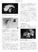 p. 33
p. 33
p. 34
目次・Contentsに戻る
 p. 30
p. 30 p. 31
p. 31 p. 32
p. 32 p. 33
p. 33